
Alannah Pearson, PhD Candidate at Australian National University. Department of Biological Anthropology
About the Researcher
Alannah Pearson is a PhD candidate in Biological Anthropology with a background in evolutionary theory and both human and non-human primate evolution. She also holds a Master of Philosophy (2015) in non-human and human greater and lesser ape cranial morphology and phylogenetic methods.
She has been an invited researcher at the Centro Nacional de Investigación sobre la Evolución Humana (CENIEH) to the hominins Paleonuerology lab under the supervision of Dr Emiliano Burner.
Summary of Project
She is studying fossil primates including the human Claude which ranges from the present day to nearly 40 million years ago. The specific issue she addressed was whether or not the size of the temporal lobe of the brain could be predicted from the cranial base where it has close spatial proximity because modern humans were believed to have larger than expected temporal lobes and when this phenomenon occurred during evolution is important for the knowledge of social behaviour which involved an increased in total brain size.
The temporal lobes of the brain are involved in a host of associated tasks including the transmission of the memory, auditory information and visual. To know whether larger temporal lobes are a phenomenon just related to modern humans or whether several other primate species have had similar transitions is important for the knowledge of primate evolution but also the human evolution within the context of the wider primate evolutionary tree.
The traditional methods to address this issue have involved using Magnetic Resonance Imaging (MRI) to determine the size of the temporal lobe in living primates including humans, or Computed Tomography (CT) scans of the cranial base to estimate the shape of fossil temporal lobe. Until my research, no attempt had been made to quantify whether the cranial base is a statistically reliable predictor of temporal lobe size in extant primates and whether that could be extrapolated into the fossil record.
The particular problem of this study is obtaining the same species used in MRI as CT and that living primates cannot be subjected to CT due to the radiation ethics. They needed to match the species in the MRI samples to those who were certified as being the same species from the skull which was CT scanned.
Approach
The approach she has taken to the solution is to compare CT from museum skull specimens which she certified species identification and matched to the MRI of the living non-human and human primates. This allowed the best opportunity that the skull and brain would be comparable.
Temporal lobe volume was taken from the human and non-human MRI and a series of 7 metrics developed to best capture the length, width and height of the temporal lobe where it is most spatially located with the inside of the cranial base.
This approach has so far proven that the temporal lobe size can be predicted from any one of the seven cranial base metrics but that some predicted temporal lobe size better in non-human primates than living humans which suggested a shift in the temporal lobe size and shape in certain areas of the cranial base.
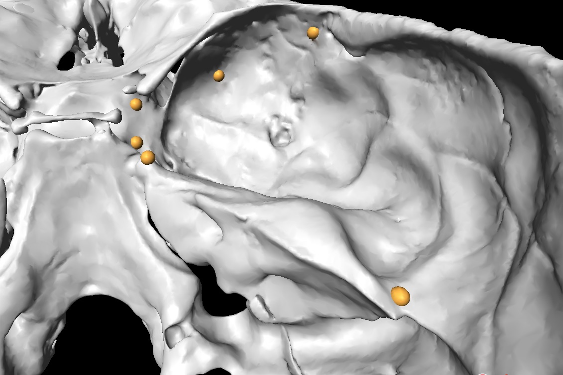
Landmark placement protocol
"My landmark placement protocol was based on the most biologically sound positions to capture the area where the temporal lobe is located most closely with the cranial base. Areas of interest included the length, width at the top, middle and further back and the location of the temporal pole plus the height at the front of the temporal lobe. These landmarks had to be repeatable on all human and non-human extant primates but also the fossil specimens which shared evolutionary branches dating back nearly 40 million years.
CheckPoint has made the collection of my data quick, easily repeatable and reliable due to the high-quality resolution and fine-tuned capabilities of the software. "
Checkpoint Testimonial
"Checkpoint has supported my research by being one of the foundation software programs that were crucial to my data collection.
The high-resolution and digital quality of the software plus the ease to manipulate large data had made checkpoint preferable to other software tools. "
Related Publications
Pearson, A., Polly, P.D., Bruner, E. (2020) Is the middle cranial fossa a reliable predictor of temporal lobe volume in extant and fossil anthropoids?
https://onlinelibrary.wiley.com/doi/10.1002/ajpa.24053
Pearson, A., P. D Polly., Bruner, E. (2021) Temporal lobe evolution in Javanese Homo erectus and African Homo ergaster: Inferences from the cranial base
https://www.sciencedirect.com/science/article/abs/pii/S104061822030450X
Pearson, A., Bruner, E., Polly, P.D. (2023) Updated imaging and phylogenetic comparative methods reassess relative temporal lobe size in anthropoids and modern humans
https://onlinelibrary.wiley.com/doi/10.1002/ajpa.24712

