If you're exploring the realm of 3D imaging analysis and visualization, understanding the capabilities of sophisticated software tools like Stratovan's Checkpoint can significantly enhance your workflow, whether you're involved in medical imaging, biological research, material science, or other fields requiring detailed 3D analysis. At its core, Checkpoint is a powerful software designed for the intricate task of visualizing, annotating, and analyzing 3D imaging data. Its utility spans several domains, primarily due to its ability to handle data from CT scans, MRI scans, and micro-CT scans, among other imaging technologies. Here's an overview of what Checkpoint offers and how it can be applied in various domains.
Key Features:
-
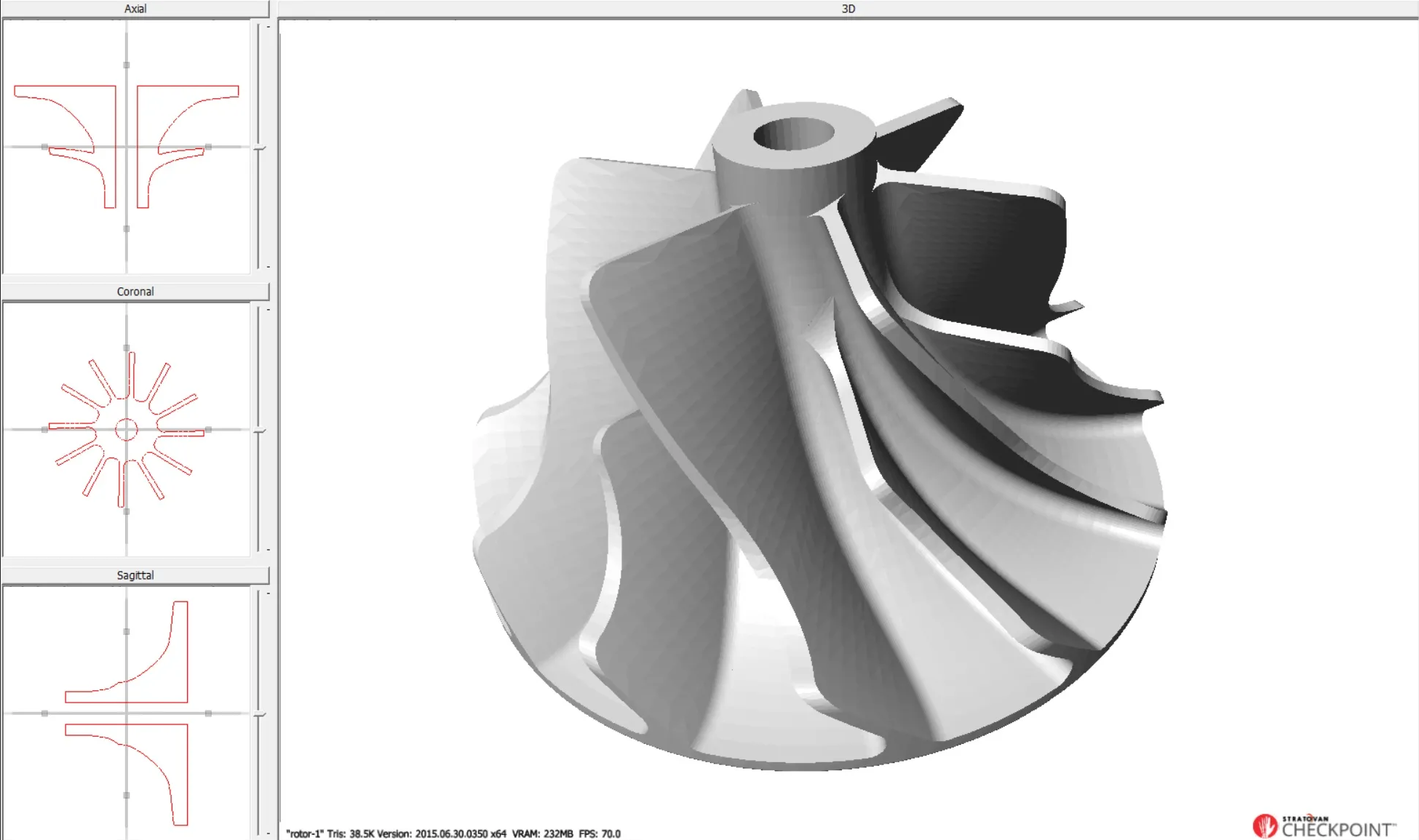
3D Visualization: Checkpoint excels in rendering 3D models from imaging data, offering interactive exploration tools that help users understand complex structures with ease. This is particularly beneficial in fields where spatial relationships and detailed structures are crucial.
-
Measurement and Analysis: The software is equipped with tools for conducting precise measurements of distances, angles, and volumes within the imaging data, facilitating rigorous quantitative analysis essential in both research and clinical settings.
-
Landmark Placement: Whether through the use of a single landmark placement point, curves, or meshes, Checkpoint supports various approaches to landmark placement to better serve the different varieties of use cases researchers and users of the tool may encounter. Very few projects, or specimens, are exactly the same....so Checkpoint needs to be robust enough to handle the subtle differences but still easy enough for users to learn and work with.
-
Collaboration: Features designed for sharing and collaborative efforts make it easier for teams to work together on complex analyses, enhancing productivity and knowledge exchange.
Applications with a Focus on Landmark Placement and Analysis:
-
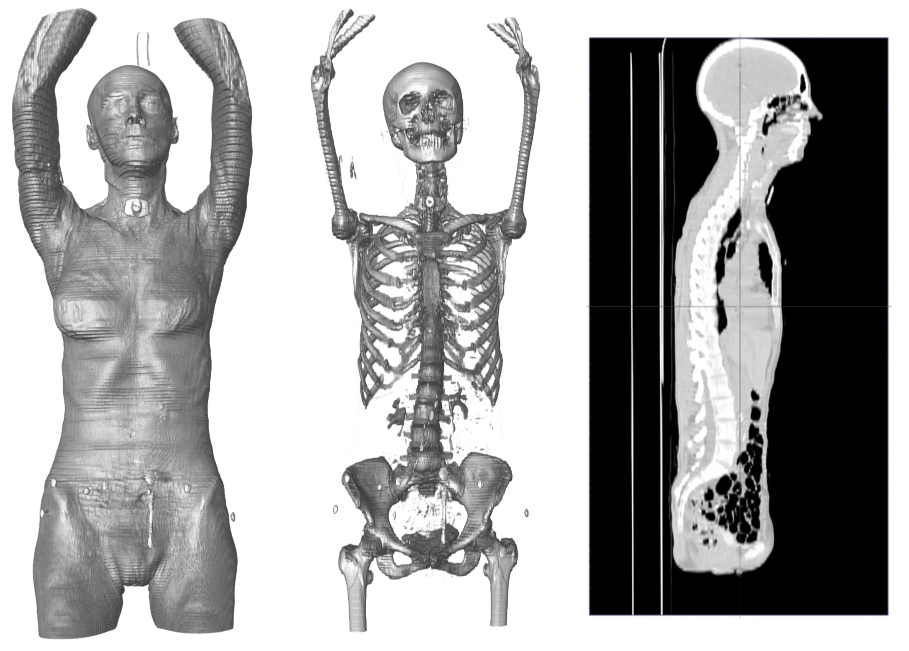
Medical Imaging: In orthopedic research, Checkpoint's landmark placement capability is crucial for comparing pre- and post-operative imagery to assess the outcomes of surgical interventions accurately. Similarly, in cardiology, placing landmarks within 3D models of the heart can help in understanding the complex geometries of heart chambers and in planning interventions for congenital heart defects.
- Example Project: A clinic utilizes Checkpoint to place landmarks around a tumor in 3D MRI scans, tracking its size and response to treatment over time, which is critical for adjusting therapy plans.
- Industry Application: In prosthetics design, technicians use landmark placement to tailor prosthetic limbs based on precise anatomical measurements, ensuring a perfect fit and optimal function for patients.
-
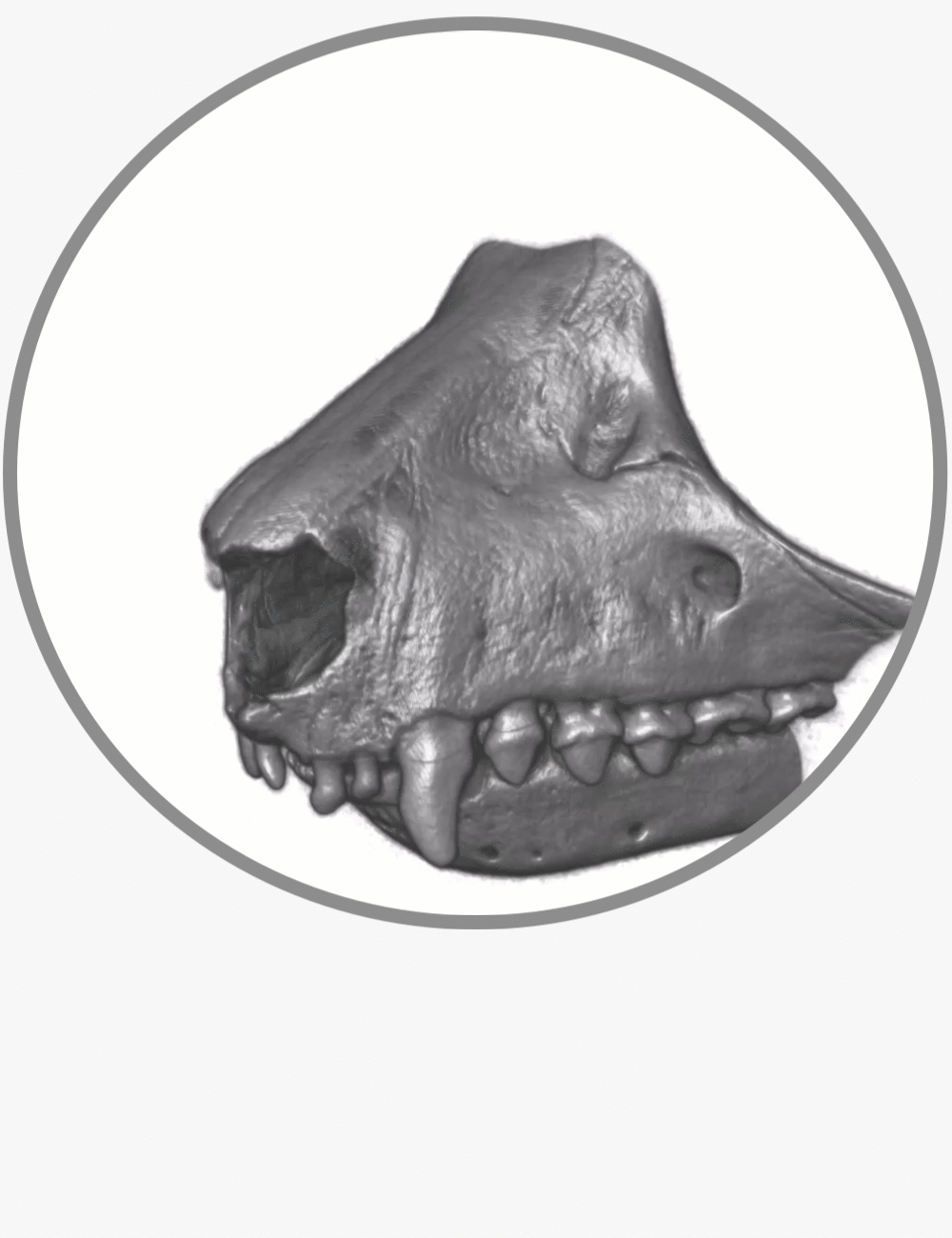
Biological Research: Landmark placement in Checkpoint enables comparative anatomy studies, allowing researchers to track developmental changes in organisms by comparing the placement of specific anatomical landmarks over time. In genetics, it aids in correlating physical trait variations with genetic differences by analyzing landmark positions in 3D models of subjects.
- Example Project: Scientists studying evolutionary biology use Checkpoint to place landmarks on 3D scans of fossilized remains, comparing them to modern species to infer evolutionary changes.
- Industry Application: In developmental biology, researchers place landmarks on 3D images of growing plant roots, analyzing how different genetic modifications affect root architecture.
-
Material Science: Checkpoint facilitates the study of materials by allowing landmarks to be placed on areas of interest within the material's structure, such as grains or pores, to quantify and analyze structural properties. This is particularly useful in assessing the quality and integrity of construction materials and in the development of new composite materials.
- Example Project: A materials science lab uses Checkpoint for placing landmarks within 3D scans of metal alloys to study the distribution and size of voids, correlating these characteristics with material strength.
- Industry Application: In semiconductor manufacturing, Checkpoint is used to place landmarks on microchip images, identifying and analyzing defect patterns to improve manufacturing processes.
-
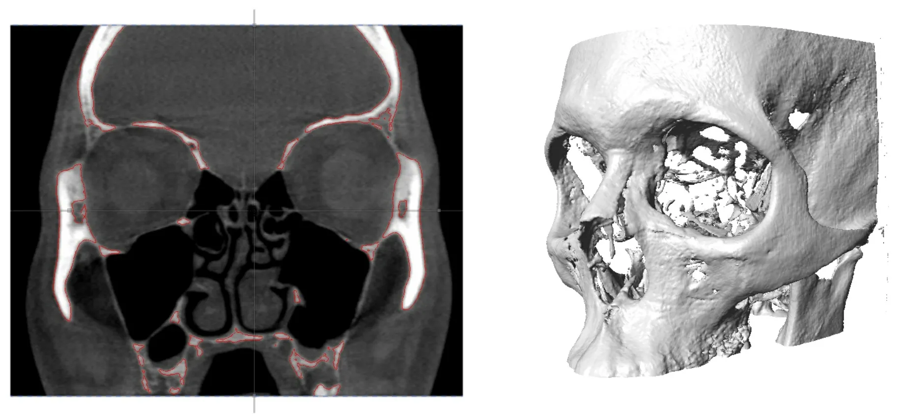
Forensics: Checkpoint's landmark placement and analysis tools are invaluable in forensic anthropology for reconstructing faces from skeletal remains. By placing landmarks at key anatomical points, forensic experts can infer the soft tissue depth and reconstruct features for identification purposes. In forensic engineering, landmarks can help in the analysis of fracture patterns or deformation in materials to understand the sequence of failures.
- Example Project: Forensic anthropologists use Checkpoint to place landmarks on a skull's surface, guiding the facial reconstruction process for unidentified remains.
- Industry Application: After a vehicle collision, forensic engineers place landmarks on deformation patterns in the vehicle's structure using Checkpoint, analyzing the data to reconstruct the accident's dynamics and identify the cause.
Through the strategic placement and analysis of landmarks, Checkpoint provides a robust framework for professionals across various industries to conduct detailed studies, enhance project outcomes, and derive meaningful insights from complex 3D data. This approach not only streamlines project workflows but also enhances the accuracy and depth of analyses, proving invaluable in advancing research, improving clinical outcomes, and solving complex forensic cases.
Why Consider Checkpoint?
For professionals and researchers seeking a user-friendly interface without sacrificing depth in 3D imaging analysis, Checkpoint stands out. Its comprehensive set of tools brings advanced imaging capabilities to your fingertips, enabling more informed decisions, whether in research, clinical applications, or education.
As you delve into the possibilities offered by software like Stratovan's Checkpoint, it's essential to stay informed about the latest advancements and how they can be tailored to fit your specific needs. Whether you're diagnosing a patient, exploring the nuances of a biological structure, analyzing material properties, or conducting forensic analysis, understanding and leveraging the power of such software can dramatically transform your approach and outcomes.
Sign up for your free trial of Checkpoint here and tell us what you think!