
The Challenge
Susan Kuzminsky, PhD, a bioarchaeologist at the Universidad Católica del Norte in Chile and the University of California at Santa Cruz, examines human skeletal remains from archaeological sites in North and South America. She wanted to find an easier, more effective, efficient and less expensive way to view, catalog and manipulate 3D images based on surface scan data generated with an HD laser scanner.
The Solution
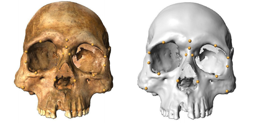
Stratovan’s Checkpoint™ for medical imaging – a comprehensive set of state-of-the-art 3D shape analysis and visualization tools, provides better landmark-based measurement capabilities for a deeper understanding of 3D anatomic structures. The solution is popular among archaeologists, but has strong applicability to healthcare (surgical planning), industrial design (biomedical/prosthetics), and many other fields.
Dr. Susan Kuzminsky, PhD, is keenly interested in technologies that can help her fellow bioarchaeologists overcome the challenges of scanning, viewing, cataloging, manipulating and comparing anatomical structures. “Human morphology changes over time and space,” says Dr. Kuzminsky. “We need tools that can more easily measure human skulls and other regions of the skeleton, and allow us to compare scans and data with international research teams.”
Dr. Kuzminsky is something of an authority on technologies for morphological measurements. She recently published a study in a journal exploring the potential of 3D laser surface scanning systems. “When 3D scanning technology came along, it was less expensive than CT machines, more portable than digital microscribes, and easier to use than some types of old school calipers. But, when we collected data around the world it was hard to replicate methods or reexamine a skeleton. Having to revisit a museum every time we wanted to reexamine a skeletal collection was labor-intensive and costly,” she states. “Other types of equipment, such as Digital Microscribes and CT imaging machines, are usually stationary. In addition, high resolution 3D images created from CT and MRI equipment can be difficult to analyze offsite because the files have to be opened in expensive software programs that require specialized training to operate.”
Scholarly Study Unearths Helpful 3D Imaging Software
In her study, Dr. Kuzminsky concluded that 3D laser scanning technology is superior to older methods used by biological anthropologists. During her testing of 3D shape analysis and visualization tools she found one solution – Stratovan Checkpoint™ for medical imaging – that offers significant advantages for archaeologists, forensic anthropologists and museum conservationists. She began using Checkpoint in her own research, and believes the solution can benefit researchers in orthopedics, industrial design, surgical planning and other fields too.
Stratovan Checkpoint is a set of state-of-the-art 3D shape analysis and visualization tools. It gives researchers better landmark-based tools that facilitate a deeper understanding of 3D anatomic structures. It provides 3D views of CT, MRI, PET and other 3D scans from a variety of modalities – including 3D surface scans – and enables the efficient collection of thousands of landmark points to provide precise analyses of complex 3D shapes.
“Several conventional methods exist for 3D shape analysis and visualization, but many of the software packages designed to be used with them are time-consuming to learn. Conventional spatial analysis systems require multiple software to download background and image files,” says Dr. Kuzminsky. “I like Stratovan Checkpoint because I can take screenshots of an image, record points on the specimen, calculate surface area and volume, and make precise measurements to more easily – and accurately – collect, analyze and use data – all with one system from one central location.”
Digging New Capabilities for Research
Dr. Kuzminsky is using Stratovan Checkpoint to study biological craniofacial variation in the early humans who peopled the New World. She is also using 3D methods and Checkpoint for bio-cultural studies of the standardization of cranial vault modification, which was a head-shaping practice common among prehistoric Andean communities. And, she used 3D models, measurements and cranial landmarks recorded in Checkpoint to analyze skeletal variation for forensic research as well.
Checkpoint enables the collection of dense sets of data points on 3D structures so researchers like Dr. Kuzminsky can more easily analyze shapes using landmarks. It provides full 3D reconstruction to easily slice through orthogonal and oblique views. It can load both surface meshes and volumetric DICOM scans, and extract multiple surfaces from a DICOM volume. A custom tool in Checkpoint analyzes joint surfaces for congruence and/or changes due to treatment or disease. It gives researchers and clinicians a one-stop tool to efficiently do precise analyses of complex 3D shapes.
The comprehensive features of Checkpoint are bringing new efficiencies to Dr. Kuzminsky’s work. “I can record data and examine the morphology of human skeletons in a much more systematic way than I could before,” she says. “Checkpoint eliminates the need to continuously go out to get data on objects we want to study, which saves time and money. In addition, since we have the 3D data, we don’t have to drop a specimen out of our sample if we make a mistake gathering information; now we can open up the file to find and correct the error in our lab.”
Endless Applications Beyond Archeology
Beyond bioarchaeology, Dr. Kuzminsky envisions applications for Checkpoint in many other industries. “Anyone working with bones in healthcare, or someone analyzing morphological differences for non-human skeletons for shape changes and variations, or even an archaeologist seeking to scan a ceramic vessel, can benefit from using Checkpoint,” she says. “It helps people open and use data sets in ways they could never do before. Researchers can share files, which expands our ability to access data sets that are shared by research teams worldwide. Documenting collections using nondestructive methods is also important in archaeology for museum stewardship and conservation. The technology also allows affordable access to collections for students, and for those in many disciplines.”
Potential use cases for Checkpoint include facilitating a biomedical engineer’s design of a new prosthetics device, or to help an orthopedic surgeon study and compare congenital defects in patients. Checkpoint could enable an orthopedist to create a bone database to develop standards based on thousands of scans, then easily compare a patient’s scans with those in the database to measure deviations.
Other uses include enabling maxillofacial surgeons to look at 3D scans of peoples’ faces to do deformity analysis on a patient, or to help plan a surgery. Checkpoint can also allow a healthcare professional to review a CT scan of a patient with a congenital bone issue. The software’s forensic applications are numerous as well. For example, Checkpoint can easily analyze bones from a criminal investigation, or accurately identify a victim from 3D bone and skull records. Checkpoint delivers the capability to collect the 3D landmark points that facilitate anatomic and shape analysis research in scores of different fields.
Importantly, Stratovan Checkpoint is extremely simple to use. “Checkpoint is very user-friendly and easy to teach to students,” says Dr. Kuzminsky. “Previously, this type of software was very hard to use and it had a steep learning curve, but it only takes about 15 minutes to get students up to speed on Checkpoint.”
Stratovan Checkpoint helps researchers in a myriad of fields more easily collect, analyze, manipulate and share data and images. This saves them time and money, and enables more accuracy and efficiencies in their work.

