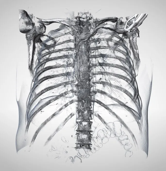
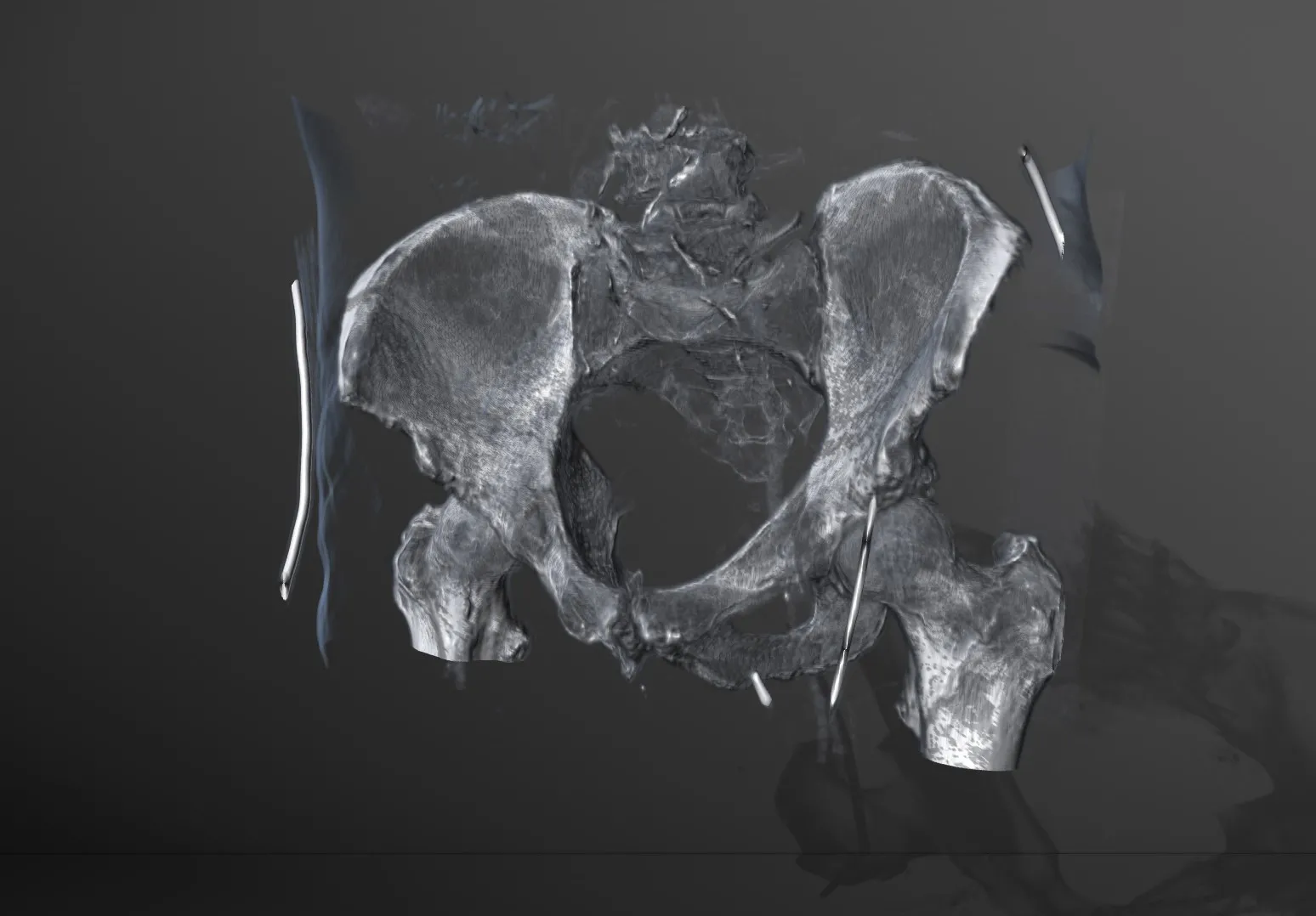
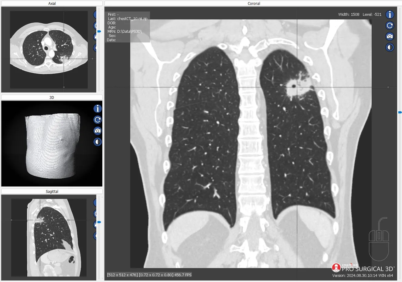
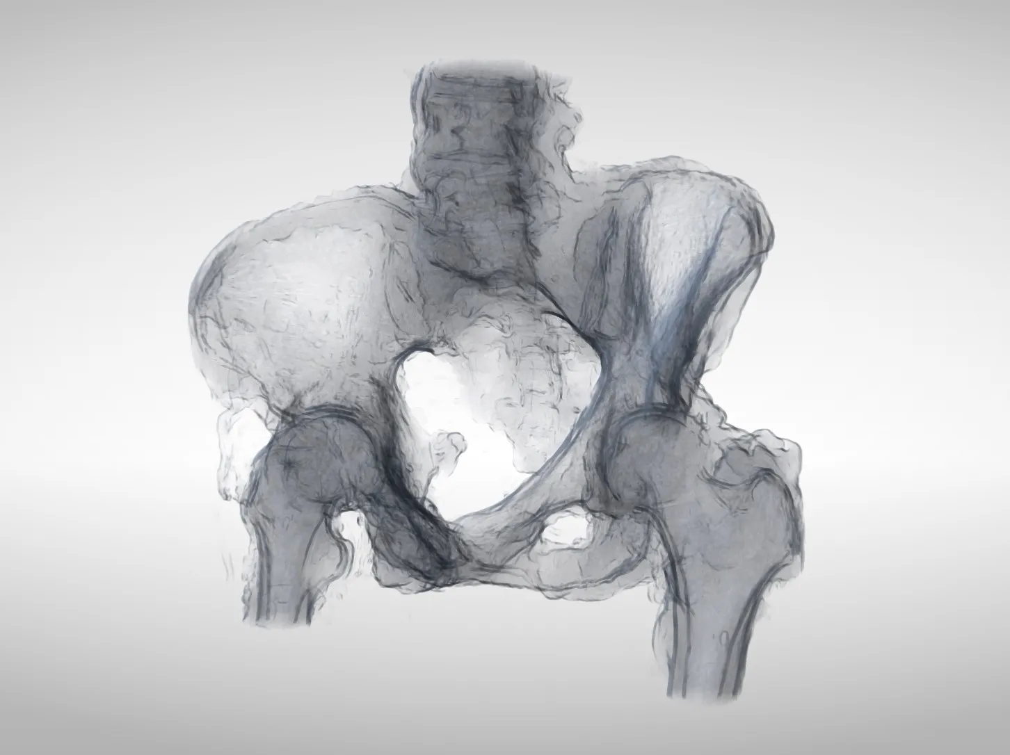
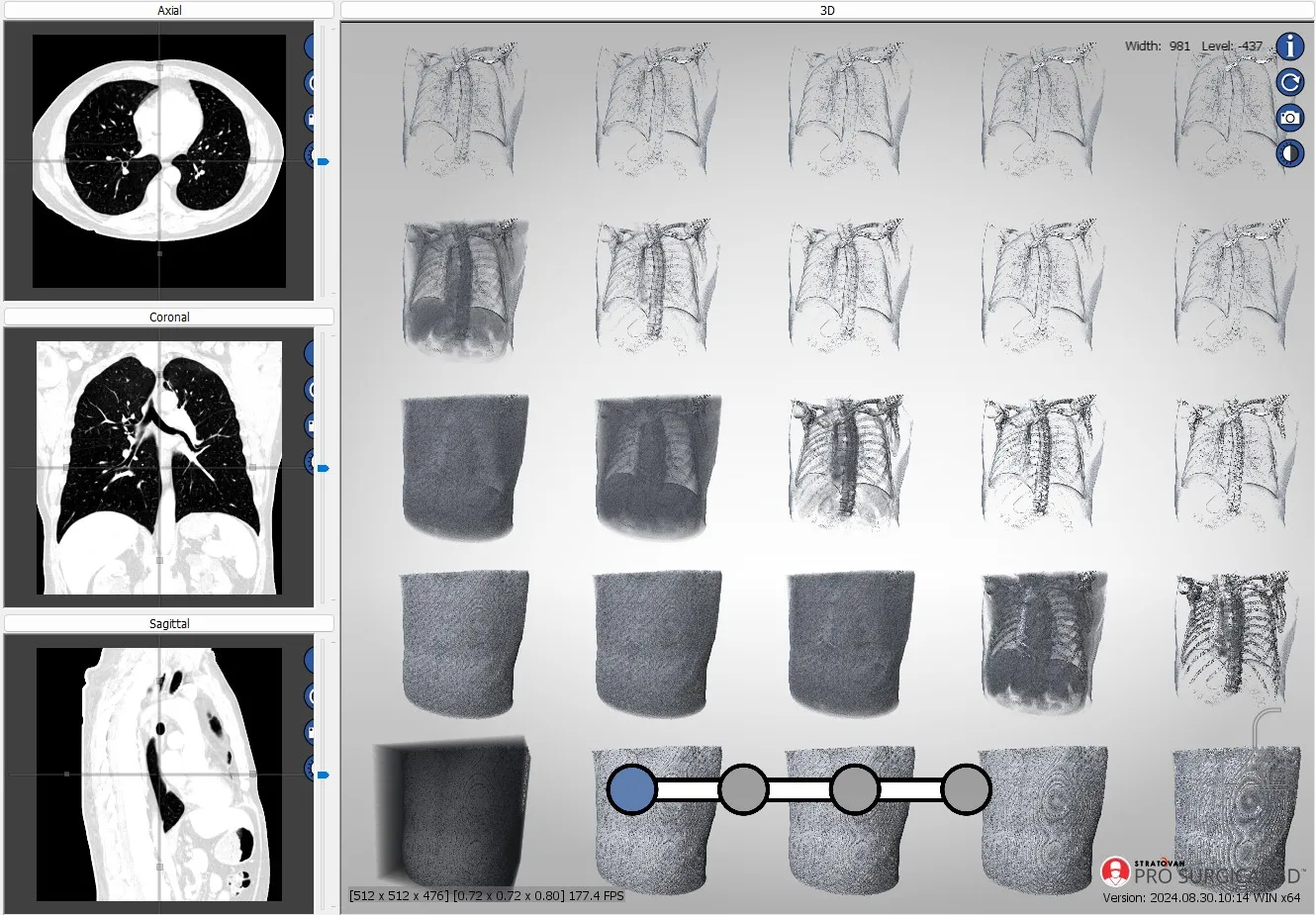
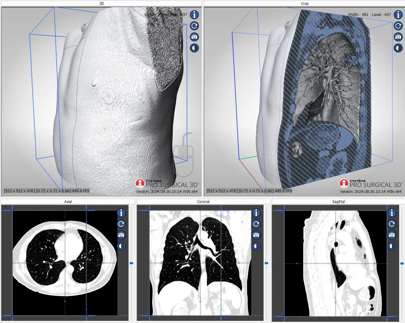
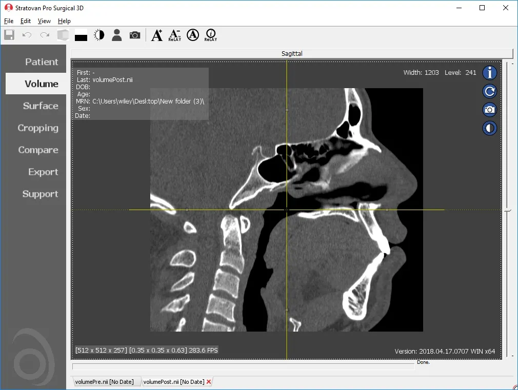
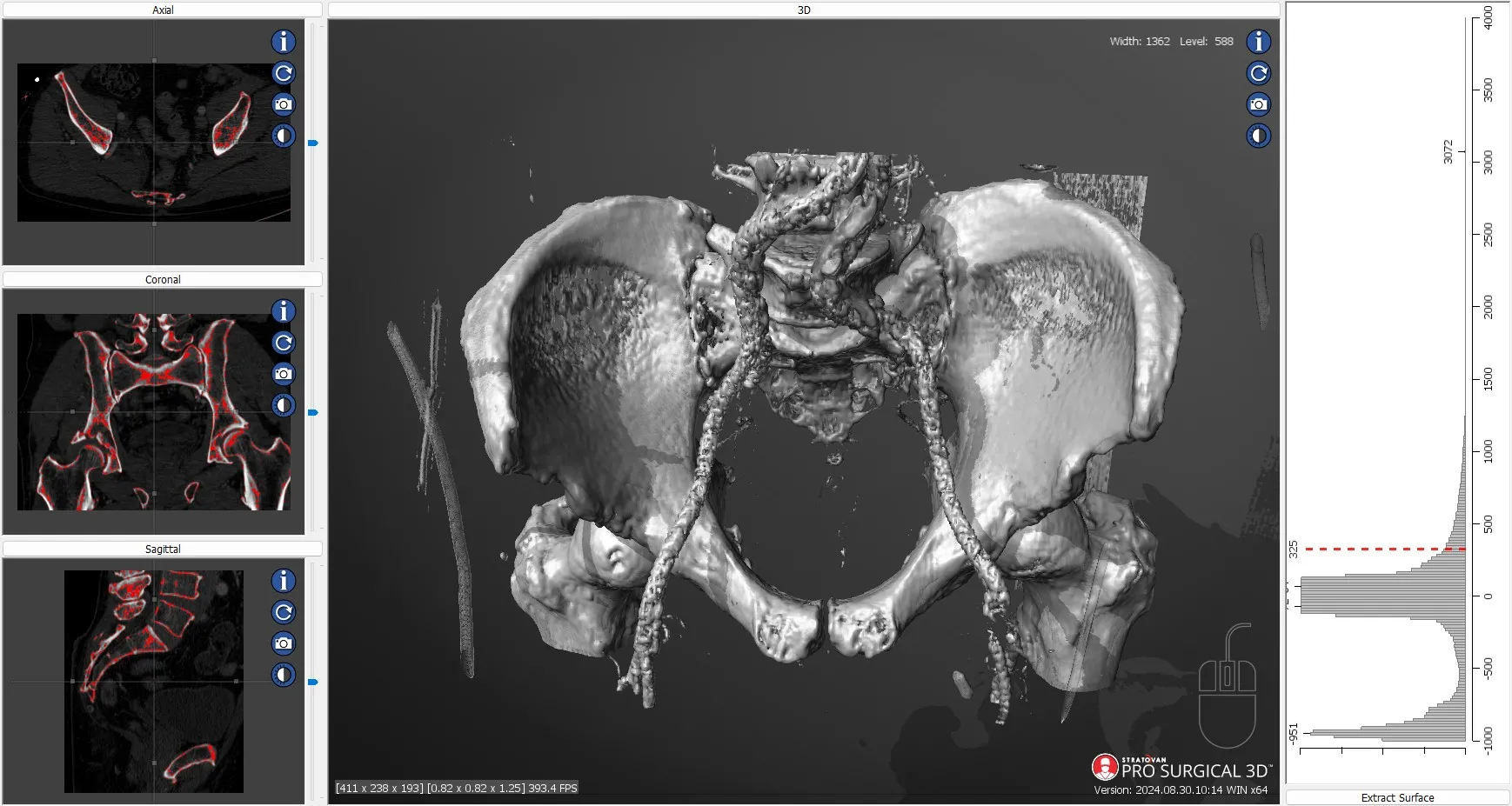
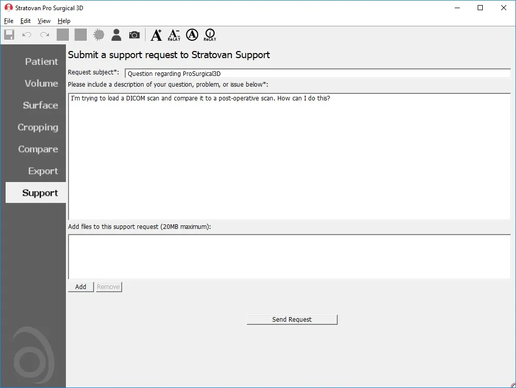
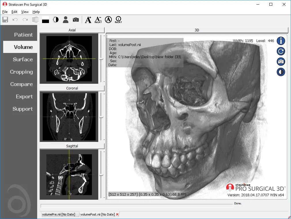
How Pro Surgical 3D benefits us all

Everyone – including surgeons, patients and their loved ones – benefits from being better informed by the wealth of information buried within CT and MRI scans.

benefits_patients
Benefits to patients

Pro Surgical 3D allows for a better understanding of a condition, disease or diagnosis, a clearer understanding of viable treatment options, a better grasp of treatment progress by comparison over time and helps to build trust and understanding with caregivers.

benefits_surgeons
For surgeons and physicians

Pro Surgical 3D is very fast and easy to use, works efficiently with patient’s DICOM scans from outside your organization, quickly loads patient scans and gives direct access from/to your PACS. Offers traditional multi-planar slicing and high-quality and fast 3D reconstruction, rendering and more…

Pro Surgical 3D brings with it a robust and detailed feature set

doctor and person looking at imaging
doctor and child hands doing a high five
doctor with child patient
  • Load DICOM data directly from PACS, CD/DVD, USB, and local computer.
  • Upload DICOM data to your PACS directly from referral patient CD/DVD and local computer.
  • Easy to use patient search to locate patient data on your PACS.
  • Loads common non-DICOM file formats: NifTi (.nii), Visualization Toolkit (.vtk), and ANALYZE (.hdr).
  • Designed mainly for CT and MR DICOM modalities.
  • Performs 3D reconstruction and volume rendering.
  • Multi-planar slicing.
  • Oblique slicing.
  • Instant and interactive surface extraction and export to STL and PLY formats.
  • Easily anonymize and de-identify patient scans.
  • Window/level (brightness and contrast) presets.
  • Screen capture.
  • Axis-aligned cropping with context.
  • Side-by-side comparative assessment for pre- and post-operative scans.
  • Integrated customer support portal.
  • and more…

Looking for 3D Image Segmentation and Annotation Labeling?

Create 3D medical image segmentation easily and in 3D. Label anatomy, create training databases for your Artificial Intelligence training process, find errors, improve ground-truth operator performance and save time and money.

3D image segmentation and annotation tools that save your team time

Segment Tab within Pro Surgical 3D Software

Easy to use: Integrated 3D image segmentation directly within Pro Surgical 3D. Get started today with delineating anatomy to create training databases for AI diagnostics, detection, and treatment planning.

World class: Tools that delineate the objects you need. Manual tools for precise voxel-by-voxel correction. Automatic AI-based image segmentation that pulls out most complex anatomy. Threshold-based tools complete the offering to give you the best tools for the job.

Segmentation quality assurance analysis using statistical outliers

Quality Assurance: How many errors are in your training database? Use the integrated quality assurance tools to find outliers. Easily load outlier scans and objects for review and fix them on the fly. Improve your final AI models by eliminating errors in your training database. You can do better.

Consistent: Labeling of annotation attributes of your objects. Create well-defined attributes that your team can use consistently across training databases. Easily change annotation attributes by batch editing across your entire training database.

product_carousel-maxillo_2
Maxillo
Maxillo

Maxillo is an orbital volume computation application for use by otolaryngologists, ENTs, plastic surgeons, craniofacial surgeons, and maxillofacial researchers. It enables the use of computed tomography (CT) scans of the orbital area to be analyzed as part of diagnostics, pre-surgical planning and post-surgical analysis method.