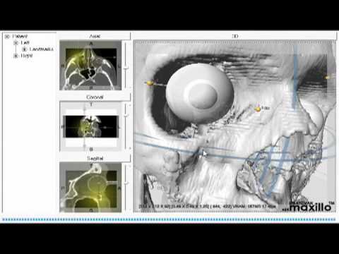
Strong, E. Bradley M.D., Fuller, Scott C. M.D., Chahal, Harinder S. M.D. (2013). Computer-Aided Analyis of Orbital Volume: A Novel Technique, Ophthalmic Plastic & Reconstructive Surgery, 29(1), 1-5.
Pham D.L., XU C.Y., and Prince J.L., (2000). A survey of current methods in medical image segmentations, Annu, Rev. Biomed. Eng., 2, 315-337.
Ramieri G, Spada M.C., Bianchi S.D., and Berrone S., (2000). Dimensions and volumes of the orbit and orbital fat in posttraumatic enophthalmus, Dentomaxillofacial Radiol., 29, 302–311.
Pohle R., and Toennies K.D., (2001). Segmentation of medical images using adaptive region growing, Proc. SPIE Med. Imag.: Image Process., 4322, 1337-1346.
Strong, E. Bradley M.D., Fuller, Scott C. M.D., Wiley, David F. PhD, Zumbansen, Janina M.D., Wilson, M. D. PhD, and Metzger, Marc C. MD, DMD, PhD, (2013). “Preformed vs Intraoperative Bending of Titanium Mesh for Orbital Reconstruction, Otolaryngology- Head and Neck Surgery, 149, 60-66.
Supporting:
Cornelius CP, Mayer P, Ehrenfeld M, and Metzger MC, (2014). The orbits--anatomical features in view of innovative surgical methods, Facial Plastic Surgery, 30, 487-508.
Zhang Z, Zhang Y, He Y, An J., Zwahlen RA, (2012). Correlation between volume of herniated orbital contents and the amount of enophthalmos in orbital floor and wall fractures. J Oral Maxillofac Surg., 70, 68-73.
Zhang ZY, He Y, Zhang Y, An JG, (2010) Evaluation of the application of computer-aided shape-adapted fabricated titanium mesh for mirroring-reconstructing orbital walls in cases of late post-traumatic enophthalmos. J Oral Maxillofac Surg., 68, 2070-2075.
Farkas LG, Posnick JC, Hreczko TM, Pron GE, (1992). Growth patterns in the orbital region: a morphometric study. Cleft Palate Craniofac J., 29, 315-318.
Karmese, Mehtap, Akdağ Osman, Selimoğlu, Muhammed Nebil, Koplay, Tugba Gun, Bakbak, Berker, Koplay, Mustafa, and Tosun, Zekeriya, (2015). Analysis of unilateral complex orbital fractures with a new treatment algorithm, Journal of Plastic Surgery and Hand Surgery, 49, 121-126.
Osaki TH, Fay A, Mehta M, Nallasamy N, Waner M, De Castro DK, (2013). Orbital Development as a Function of Age in Indigenous North American Skeletons, Ophthal Plast Reconstr Surg., 2, 131-136.
Ellis E 3rd, (2012). Reconstruction of orbital floor defects, Journal of Oral and Maxillofacial Surgery : Official Journal of the American Association of Oral and Maxillofacial Surgeons, 70, 2255.
Baino F., (2011). Biomaterials and implants for orbital repair, Acta Biomaterialia, 7, 3248-3266.
Ellis E 3rd, (2014). Surgical approaches to the orbit in primary and secondary reconstruction, Facial Plastic Surgery, 30, 537-544.
Ellis E 3rd, Perez D, (2014). An algorithm for the treatment of isolated zygomatico-orbital fractures, Journal of Oral and Maxillofacial Surgery : Official Journal of the American Association of Oral and Maxillofacial Surgeons, 72, 1975-1983.
Nysjö, Johan, (2011). Orbit Segmentation for Cranio- Maxillofacial Surgery Planning, Upsalla University - Department of Information Technology, 1-59.
Song SY, Hong JW, Roh TS, Kim YO, Kim DW, Park BY, (2009). Volume and distances of the maxillary sinus in craniofacial deformities with midfacial hypoplasia, Otolaryngol Head Neck Surg., 141, 614-620.
Lieger, O., Richards, R, Liu, M, Lloyd T, ( 2010). Computer-assisted design and manufacture of implants in the late reconstruction of extensive orbital fractures, Arch Facial Plast Surg., 3, 186-191.
Forbes G, Gehring D.G., Gorman C.A., Brennan M.D., and Jackson I.T., (1985). Volume measurements of normal orbital structures by computed tomographic analysis quantitative volumetric assessment of orbital soft tissue, AJNR, 6, 419–424.
Krahe T, Schlolaut K.H., and Poss T, et al. (1989). Computed tomographic volumetry of the orbit in endocrine orbitopathy, Rofo., 151, 597–601.
R A Nugent, R.A., Belkin, R.I., Neigel, J.M., Roodman, J., Robertson, W.D., Spinelli, J. and Graeb, D.A., (1990). Graves orbitopathy: correlation of CT and clinical findings, Radiology, 177, 675-682.
Zonneveld F.W, Koornneef L., and Wittebol-Post D. (1991). Quantitative volumetric assessment of orbital soft tissue, Computer Assisted Radiology, 181-186.
Lutzemberger L, and Salvetti O., (1998). Volumetric analysis of CT orbital images, Med Biol Eng Comput. 36, 661–666.
Pham D.L., XU C.Y., and Prince J.L., (2000). A survey of current methods in medical image segmentations, Annu, Rev. Biomed. Eng., 2, 315-337.
Ramieri G, Spada M.C., Bianchi S.D., and Berrone S., (2000). Dimensions and volumes of the orbit and orbital fat in posttraumatic enophthalmus, Dentomaxillofacial Radiol., 29, 302–311.
Pohle R., and Toennies K.D., (2001). Segmentation of medical images using adaptive region growing, Proc. SPIE Med. Imag.: Image Process., 4322, 1337-1346.
Yushkevich P.A., Piven J., Hazlett H.C., Smith R.G.,, Ho S., Gee J.J., and Gerig G., (2006). User-guided 3-D active contour segmentation of anatomical structures: Significantly improved efficiency and reliability,
NeuroImage, 31, 1116-1128.
Bijlsma W.R., and Mourits M.P. (2006). Radiologic measurements of extraocular muscle volumes in patients with Graves’ orbitopathy: a review and guideline, Orbit. 25, 83–91.
Regensburg, N.I., Wiersinga, W.M., van Velthoven, M.E.J., Berendschot, T.T.J.M., Zonneveld, F.W., Baldeschi, L., Saeed, P. and Mourits, M.P., (2009). Age and gender-specific reference values of orbital fat and muscle volumes in Caucasians, British Journal of Ophthalmology, doi:10.1136/bjo.2009.161372.
Alsuhaibani, A.H., Carter, K.D., Policeni, B. and Nerad, J.A. (2010). Effect of orbital bony decompression for Graves' orbitopathy on the volume of extraocular muscles, British Journal of Ophthalmology, 95, 1255-1258.
Weaver, A.A., Loftis, K.L., Tan, J.C., Duma, S.M., and Stitzel, J.D., (2010). CT Based Three-Dimensional Measurement of Orbit and Eye Anthropometry, Investigative Ophthalmology & Visual Science, 51, 4892-4897.